Granulomatous Disease Lung Nodules
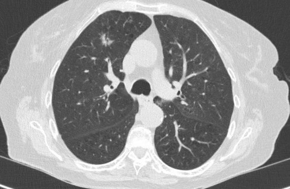
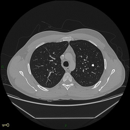
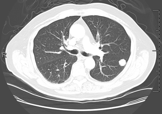
Granulomatous disease lung nodules. Typically granulomas are noncancerous benign. A granuloma is a small area of inflammation. One or more lung nodules can be an incidental finding found in up to 02 of chest X-rays and around 1 of CT scans.
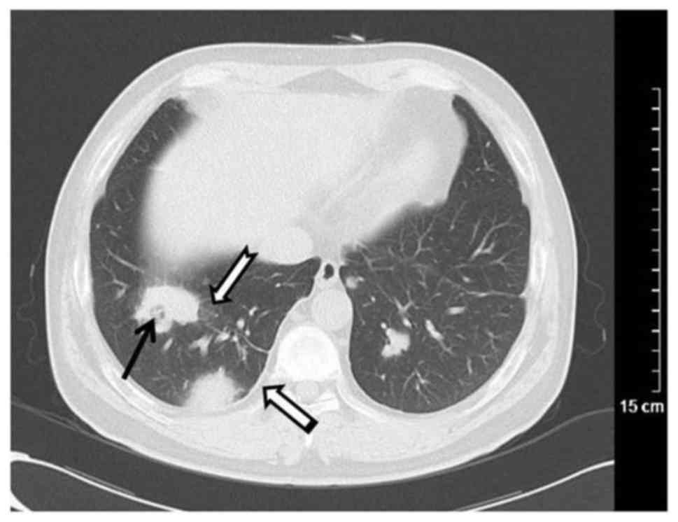
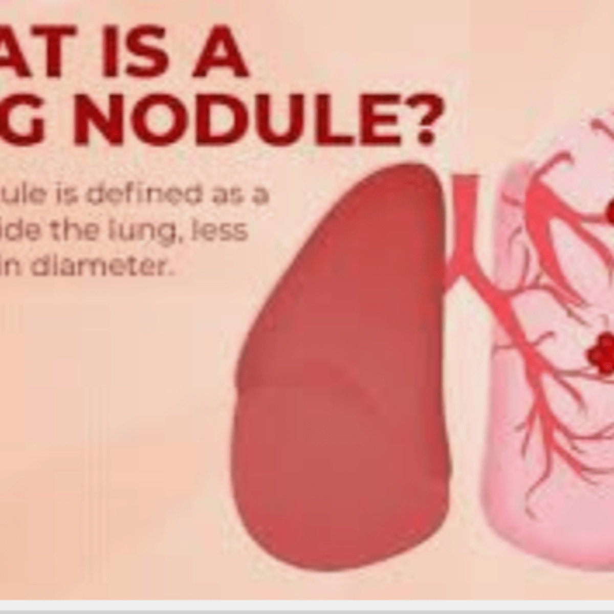
A lung nodule or pulmonary nodule is a relatively small focal density in the lung. Pulmonary non-tuberculous mycobacterial infection. In some series of solitary pulmonary nodules in endemic areas coccidioidal granulomas are more common than malignancies.
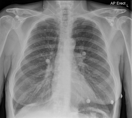
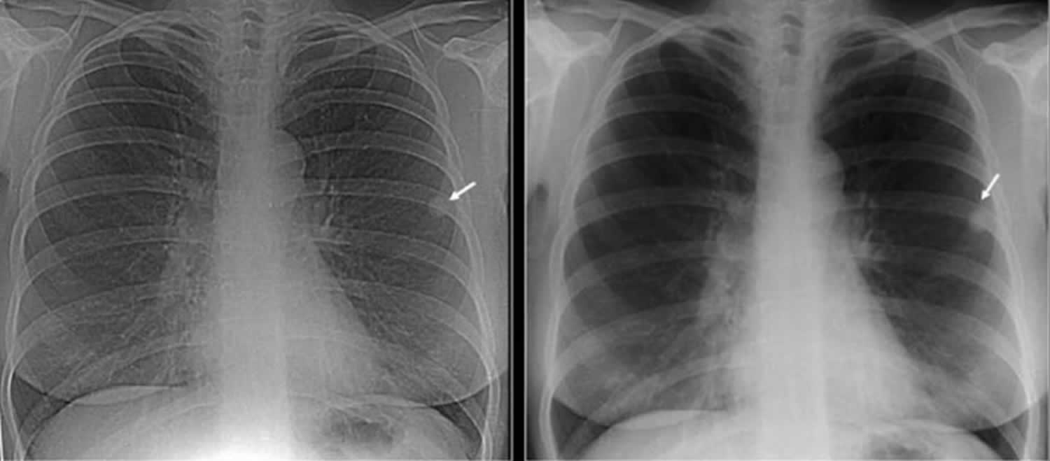
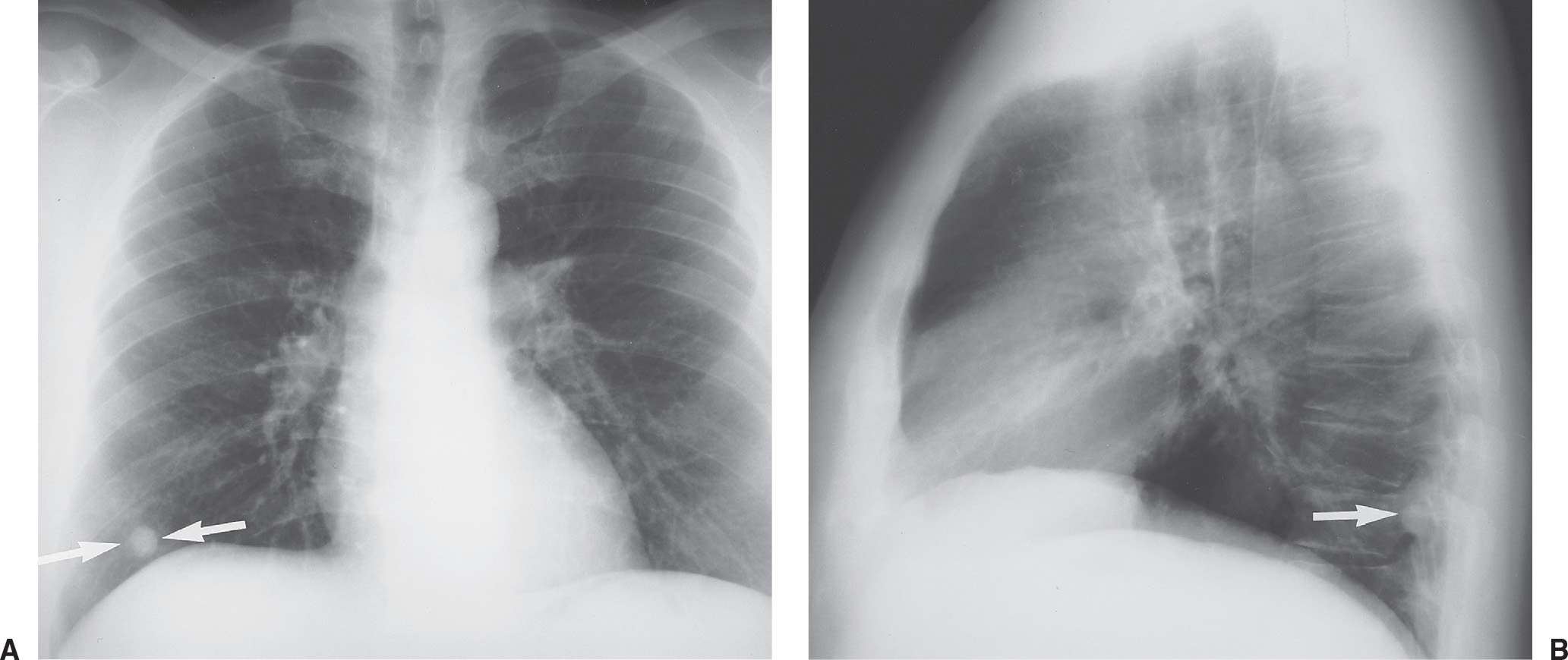
Dystrophic calcification follows caseation necrosis or fibrosis. A solitary pulmonary nodule or coin lesion is a mass in the lung smaller than 3 centimeters in diameter. Granulomas are often found incidentally on an X-ray or other imaging test done for a different reason.
RA primarily affects your joints but it can cause lung granulomas also. Neurofibromas are a type of noncancerous neoplasm. They are typically detected through an x-ray or CT scan of the lungs and are often initially discovered accidentally when a patient receives one of these scans for a separate concern.
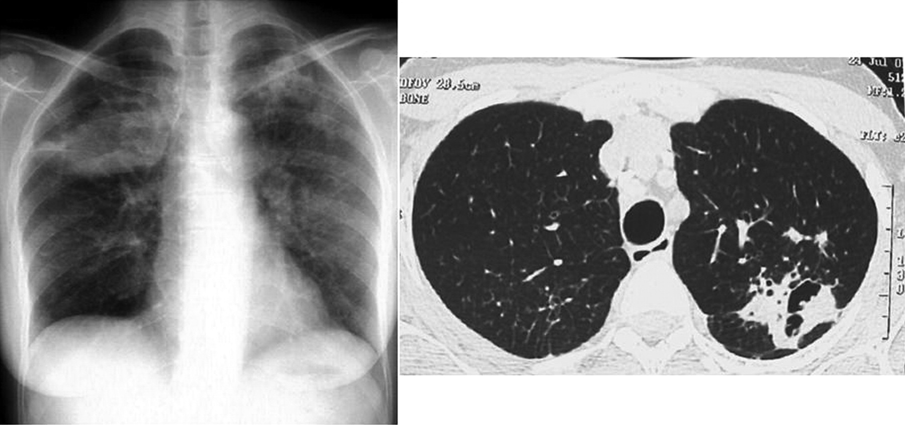
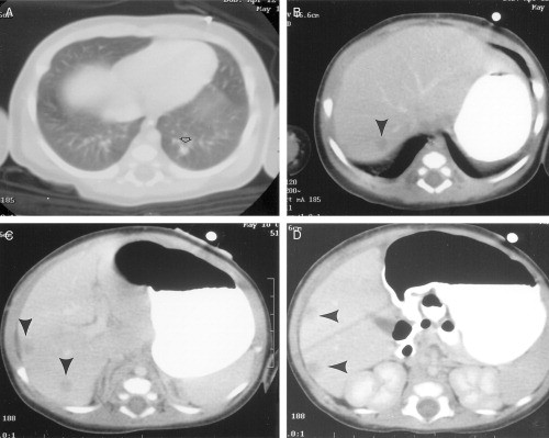
Differential diagnosis is challenging and includes both infectious mycobacteria and fungi and noninfectious lung diseases sarcoidosis necrotising sarcoid granulomatosis hypersensitivity pneumonitis hot tub lung berylliosis granulomatosis with. Lung nodule is used to refer to round or oval lesions on the lung that are 3 cm in diameter or less. The pulmonary radiologic findings of chronic granulomatous disease include consolidation nodules areas of scarring traction bronchiectasis emphysema air trapping mediastinal and hilar lymphadenopathy pulmonary artery enlargement and pleural effusion.
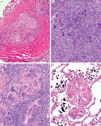
When an infection or illness inflames lung tissue a small clump of cells granuloma can form. Granulomatous lung disease refers to a broad group of infectious and non-infectious conditions characterized by the formation of granulomas. Healed andor contained infections can cause lung nodules or cavities that mimic malignancy analogous to histoplasmomas see Clinical and Radiologic Findings section of histoplasmosis.
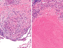
The nodular lung lesions corresponded to typical histologically proved rheumatoid nodules in 4 cases and aseptic and noncaseating granulomatous lung inflammation in 3 cases. These cases were mainly observed with etanercept 611 and occurred in patients receiving associated disease-modifying antirheumatic drugs DMARD such as methotrexate or leflunomide in 8 cases.
Dystrophic calcification follows caseation necrosis or fibrosis.
Noninfectious diagnoses 15 of 79 were rheumatoid nodule five granulomatosis with polyangiitis Wegener five sarcoidosis four and chronic granulomatous disease one. RA primarily affects your joints but it can cause lung granulomas also. Precise clinical evaluation laboratory testing pulmonary function testing radiological imaging including high-resolution computed tomography and often histopathological assessment contribute to make a confident diagnosis of granulomatous lung diseases. Cocci outbreaks observed in pacific northwest May be clinically significant in some patients especially the. Many cases remained unexplained even after extensive review 52 of 131 40. Calcified nodules following infections are well defined and often measure 2-5 mm in diameter. Pulmonary non-tuberculous mycobacterial infection. To evaluate combined PET-computed tomography CT criteria for differentiating between granulomatous disease GD and malignancy CA in oncologic PET-CT studies. To estimate the diagnostic accuracy of signal intensity of the lesion-to-spinal cord ratio LSR and apparent diffusion coefficient ADC in diffusion-weighted DW magnetic resonance imaging of pulmonary nodules suspicious for lung cancer in granulomatous lung disease-endemic regions.
Calcified granulomas can also form. Typically granulomas are noncancerous benign. A solitary pulmonary nodule or coin lesion is a mass in the lung smaller than 3 centimeters in diameter. The nodular lung lesions corresponded to typical histologically proved rheumatoid nodules in 4 cases and aseptic and noncaseating granulomatous lung inflammation in 3 cases. Lung nodule is used to refer to round or oval lesions on the lung that are 3 cm in diameter or less. Healed andor contained infections can cause lung nodules or cavities that mimic malignancy analogous to histoplasmomas see Clinical and Radiologic Findings section of histoplasmosis. A granuloma is a small area of inflammation.









/iStock-12102066521-42ae870d7a904f0bba0911fbda5ec64a.jpg)








:max_bytes(150000):strip_icc()/lung-nodules-symptoms-causes-and-diagnosis-2249304_final11-5b44dd5cc9e77c003735e393.png)




Post a Comment for "Granulomatous Disease Lung Nodules"